THE IMPACT OF POSTERIOR STAPHYLOMA ON CLINICAL OUTCOMES OF HIGH MYOPIC MACULAR HOLE: A Retrospective Cohort Study.
Zheng et al.
Retina·Mar 1, 2026
In high myopic macular holes, inverted ILM flap technique achieved superior closure rates compared to ILM peeling across different posterior staphyloma types and grades. Staphyloma morphology significantly influences surgical outcomes and vascular recovery patterns.
Impact of Phacoemulsification on Vitreomacular Traction Release and Complications.
Bala et al.
Am J Ophthalmol·Mar 1, 2026
This retrospective study of 310 eyes found that phacoemulsification does not significantly increase the likelihood or speed of vitreomacular traction (VMT) release, with intrinsic patient factors being more predictive of outcomes.
Long Term Anatomic and Visual Outcomes of Surgery for Rhegmatogenous Retinal Detachment in Young Adult Patients.
Oh et al.
Ophthalmol Retina·Feb 26, 2026
Primary scleral buckle repair for rhegmatogenous retinal detachment in young adults achieves 80.3% single-surgery success at 12 months with favorable long-term visual outcomes, though 26.9% require fellow-eye intervention during follow-up.
Comparing the Vitreous Floaters Functional Questionnaire to the VFQ-25 in Vision Degrading Myodesopsia from Vitreous Floaters.
Sebag et al.
Am J Ophthalmol·Feb 24, 2026
The Vitreous Floater Functional Questionnaire (VFFQ-23) significantly outperformed the NEI VFQ-25 in predicting surgical decision-making and measuring post-operative improvement in patients with vision-degrading myodesopsia from vitreous floaters.
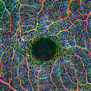
Elevated Retinal Neovascularization on Widefield Optical Coherence Tomography Angiography Predicts Complications in High-Risk Proliferative Diabetic Retinopathy.
Wu et al.
Am J Ophthalmol·Mar 1, 2026
Widefield SS-OCTA-derived metrics of retinal neovascularization burden, particularly elevated RNV lesions separated from the internal limiting membrane, predict vision-threatening complications in high-risk proliferative diabetic retinopathy with high accuracy.
Association of Patient Age with Response to Anti-Vascular Endothelial Growth Factor Agents for Treatment of Diabetic Macular Edema.
Liu et al.
Retina·Mar 1, 2026
Anti-VEGF treatment response for diabetic macular edema significantly decreases with patient age, with older patients showing less visual acuity improvement and higher rates of weak treatment response over 2 years.
CORRELATION OF RETINAL IMAGING UTILIZATION WITH ANTI-VASCULAR ENDOTHELIAL GROWTH FACTOR AND PANRETINAL PHOTOCOAGULATION USAGE FROM 2013 TO 2021.
Sheth et al.
Retina·Mar 1, 2026
OCT utilization increased significantly from 2013-2021 while fluorescein angiography declined, correlating with increased anti-VEGF injections and decreased panretinal photocoagulation usage in retinal disease management.
OUTER RETINAL LAYER DETERIORATION PATTERNS IN EYES WITH DIABETIC SEROUS MACULAR DETACHMENT.
Kirik et al.
Retina·Mar 1, 2026
This retrospective study identified five distinct outer retinal layer deterioration patterns in diabetic serous macular detachment, with WEDGE pattern showing the best visual prognosis and DIFFUSE pattern having the poorest outcomes.
Expanded Field Optical Coherence Tomography Angiography Biomarkers Associated with Future Cardiovascular Disease and Mortality in Patients with Diabetic Retinopathy.
Lu et al.
Retina·Mar 1, 2026
This study found that specific OCTA biomarkers, including neovascularization vessel density and deep capillary plexus changes, are associated with increased risk of cardiovascular events and mortality in diabetic retinopathy patients.
Association Between Baseline Glycated Hemoglobin Levels and Functional and Anatomical Outcomes Following Faricimab Loading Phase in Diabetic Macular Edema.
Scampoli et al.
Retina·Feb 24, 2026
In 74 eyes with diabetic macular edema, baseline HbA1c levels showed weak correlation with visual acuity improvement but did not predict faricimab treatment response. Faricimab loading phase demonstrated significant anatomical and functional improvements regardless of glycemic control status.
This pilot study describes choroidal synphlebia, a newly recognized choroidal venous phenotype characterized by confluent venous structures ≥750 µm, predominantly associated with central serous chorioretinopathy and potentially explaining treatment-resistant cases.
A Randomized-Controlled Trial of the Efficacy, Safety and Tolerability of Intravitreal Brolucizumab in Patients With Chronic Central Serous Chorioretinopathy With Persistent Fluid in the Absence of Choroidal Neovascular Membrane-BRICS Trial Report II.
Ramamurthy et al.
Am J Ophthalmol·Mar 1, 2026
This randomized controlled trial demonstrated that intravitreal brolucizumab effectively resolves persistent subretinal fluid in chronic central serous chorioretinopathy without choroidal neovascularization, with 77% of treated eyes achieving >20% reduction in central macular thickness at 1 month.
Atrophy Advisor: A Clinical Tool for Dry Macular Degeneration With Geographic Atrophy.
Kerwin et al.
Am J Ophthalmol·Mar 1, 2026
Kerwin et al. developed Atrophy Advisor, a clinical decision tool that combines geographic atrophy progression rates and personalized lifespan estimates to guide complement factor inhibitor treatment decisions in dry AMD patients.
Basal Laminar Deposits and Pseudodrusen: Rethinking Their Role in Age-Related Macular Degeneration Progression.
Fragiotta et al.
Retina·Mar 1, 2026
Basal laminar deposits (BLamD) are consistently present in eyes with pseudodrusen and may serve as indicators of AMD severity, potentially contributing to rapidly progressive atrophy when associated with subretinal drusenoid deposits.
Retinal fingerprint pattern in central serous chorioretinopathy.
Burnasheva et al.
Retina·Mar 1, 2026
Retinal fingerprint pattern (RFP) was identified in 18.3% of central serous chorioretinopathy (CSCR) eyes and was associated with greater subretinal fluid height and significantly better anatomical outcomes including higher rates of complete fluid resolution.
DIAGNOSTIC PERFORMANCE OF MACHINE LEARNING TECHNOLOGY USING OPTICAL COHERENCE TOMOGRAPHIC IMAGE IN RETINAL DISEASES PRESENTED WITH SUBRETINAL FLUID.
Khakhai et al.
Retina·Mar 1, 2026
Machine learning analysis of OCT images achieved 87.10% accuracy in differentiating central serous chorioretinopathy, polypoidal choroidal vasculopathy, and Vogt-Koyanagi-Harada disease, with best performance using single foveal OCT scans combined with infrared photography.
A NOVEL TECHNIQUE FOR SUBRETINAL HEMATOMA MIGRATION: Directing the Lesion Toward Artificial Retinal Detachment.
Goda et al.
Retina·Mar 1, 2026
A novel surgical technique demonstrates that subretinal hematomas can be directed away from the fovea by creating an artificial retinal detachment with subretinal tPA injection at a strategic location temporal to the macula.
OPTICAL COHERENCE TOMOGRAPHY AND OPTICAL COHERENCE TOMOGRAPHY ANGIOGRAPHY BIOMARKERS FOR PREDICTING RESPONSE TO ANTI-VASCULAR ENDOTHELIAL GROWTH FACTOR THERAPY IN NEOVASCULAR AGE-RELATED MACULAR DEGENERATION.
Ucak et al.
Retina·Mar 1, 2026
Loop-shaped neovascular morphology on baseline OCTA independently predicted poor anatomical response to anti-VEGF therapy in treatment-naïve neovascular AMD patients. This biomarker may enable early risk stratification and guide personalized treatment approaches.
Quantifying Effects of Diet and Lifestyle Changes on Progression to Advanced Age and Related Macular Degeneration in High Genetic Risk Individuals.
Seddon et al.
Ophthalmology·Mar 1, 2026
Among high genetic risk patients with early/intermediate AMD, adopting healthy lifestyle behaviors could prevent 56-60% of progression to advanced AMD, with unhealthy behaviors increasing progression risk 3-5 fold.
Clinical Characteristics and Outcomes in Central Serous Chorioretinopathy with Subretinal Hyper-Reflective Material: MICRoN report 6.
Sahoo et al.
Am J Ophthalmol·Feb 26, 2026
Chronic CSCR patients with subretinal hyperreflective material (SHRM) present with worse baseline vision and have higher rates of persistent subretinal fluid and ellipsoid zone loss despite visual improvement after treatment.
Domain-Shift AI Technology for Vendor-Agnostic Multiple Macular Disease Detection From 3D OCT Scans.
Tang et al.
JAMA Ophthalmol·Feb 26, 2026
A deep learning model trained exclusively on Spectralis OCT scans demonstrated robust performance for multiple macular disease detection across different OCT vendors (Heidelberg and Zeiss) with AUROC values ranging from 0.754-0.999, achieving high negative predictive values (>97.5%) and manageable miss rates for urgent cases (6-7%).
Comparison of Initial Loading Injection Outcomes Between Aflibercept 8 mg and Faricimab in Neovascular Age-Related Macular Degeneration.
Han et al.
Retina·Feb 25, 2026
This retrospective study of 100 treatment-naïve neovascular AMD patients found that aflibercept 8 mg and faricimab achieved statistically equivalent visual and anatomical outcomes after three monthly loading injections.
SD-OCT and OCTA: distinct or additional information on neovascular activity? The COCTAEYL Study.
Amoroso et al.
Retina·Feb 24, 2026
OCTA showed poor diagnostic performance compared to SD-OCT for detecting neovascular AMD recurrence, with sensitivity of 72% but specificity of only 27%, and did not reduce unnecessary injections or change treatment decisions.
Tomographic biomarkers differ by progression risk to late macular degeneration.
Bernardi et al.
Ophthalmol Retina·Feb 24, 2026
Hyperreflective foci (HRF) and subretinal drusenoid deposits (SDD) show distinct progression patterns in AMD, with HRF increasing risk for both geographic atrophy and neovascular AMD, while SDD increases risk only for geographic atrophy.
CRVO, BRVO, retinal ischemia, macular edema from RVO
CHOROIDAL VASCULARITY INDEX, RETINAL VASCULARITY, AND HEMOGLOBIN LEVELS IN PEDIATRIC SICKLE CELL MACULOPATHY.
Chaaya et al.
Retina·Mar 1, 2026
Pediatric sickle cell disease patients showed significantly reduced choroidal vascularity index (54% vs 69% in controls) that correlated with decreased retinal vessel density and lower hemoglobin levels, suggesting choroidal ischemia contributes to sickle cell maculopathy pathogenesis.
Association between Retinal Vein Occlusion and Epiretinal Membrane Development.
Almosa et al.
Am J Ophthalmol·Feb 24, 2026
Epiretinal membrane (ERM) formation peaks within 3 months after retinal vein occlusion (RVO), with highest surgical intervention rates occurring at 1 year for BRVO and 3 years for CRVO in untreated eyes.
Natural History of CNGA1-Associated Retinitis Pigmentosa in a Large Chinese Cohort Revealing an Optimal Intervention Window.
Liu et al.
Am J Ophthalmol·Mar 1, 2026
This large Chinese cohort study of 58 CNGA1-associated retinitis pigmentosa patients identified a distinct natural history pattern with visual acuity remaining stable until age 30.7 years before declining, while visual field loss occurs earlier, suggesting an optimal intervention window between the 3rd and 4th decades of life.
Autosomal Recessive Bestrophinopathy-Phenotypic Variability, Natural History, and Genotype-Phenotype Correlations.
Bianco et al.
Am J Ophthalmol·Mar 1, 2026
Autosomal recessive bestrophinopathy shows wide phenotypic variability from mild macular involvement to severe panretinal degeneration, but visual impairment risk is primarily driven by primary angle closure rather than fundus lesion severity.
EARLY FINDINGS FROM A NATURAL HISTORY STUDY OF PATIENTS WITH THE PATHOGENIC p.Gly208Asp PRPH2 VARIANT ASSOCIATED WITH RETINAL DYSTROPHY.
AlAshwal et al.
Retina·Mar 1, 2026
This prospective study of 7 patients with the pathogenic p.Gly208Asp PRPH2 variant demonstrates significant retinal thinning and expansion of atrophic areas over 1 year, establishing multimodal imaging markers for monitoring central areolar chorioretinal dystrophy progression.
MERTK-related retinopathy presents as juvenile rod-cone dystrophy with early macular involvement, typically causing significant visual loss by teenage years with characteristic asymmetric central macular atrophy.
Progression in X-Linked Retinoschisis: A Longitudinal Study Defining Quantitative Biomarkers and Their Implications for Gene Therapy.
Wei et al.
Am J Ophthalmol·Feb 25, 2026
This longitudinal study of 107 XLRS patients demonstrates a triphasic pattern of visual decline with optimal therapeutic window in early adulthood, and validates automated cyst-cavity volume as a quantitative biomarker for monitoring disease progression.
EFFICACY AND VASCULAR REDEVELOPMENT OF ANTI-VASCULAR ENDOTHELIAL GROWTH FACTOR THERAPY FOR STAGE 2 EARLY-DIAGNOSED FAMILIAL EXUDATIVE VITREORETINOPATHY.
Ma et al.
Retina·Mar 1, 2026
Anti-VEGF therapy effectively treated 79% of stage 2 FEVR cases diagnosed under 3 months of age and promoted continued peripheral vascular growth, with most eyes showing regression of vascular activity.
Re: Barresi et al: Surgical and Observational Outcomes in Optic Pit Maculopathy: A Comparative Analysis of Pediatric and Adult Populations (Ophthalmol Retina. 2026;10:445-454).
Shukla
Ophthalmol Retina·Feb 26, 2026
Abstract not available — see full article for details.
Interpreting Imaging in the Era of Artificial Intelligence: Future Possibilities in Ocular Inflammatory Disease.
McKay et al.
Am J Ophthalmol·Mar 1, 2026
This review examines the emerging applications of artificial intelligence in uveitis diagnosis and monitoring, highlighting AI's potential to automate multimodal imaging interpretation and improve clinical efficiency in ocular inflammatory disease management.
Factors Associated with Ocular Relapse and Visual Prognosis in Sarcoid Uveitis.
Abramowicz et al.
Ophthalmology·Mar 1, 2026
In this multicenter study of 336 sarcoid uveitis patients, ocular relapses occurred in two-thirds of patients with macular edema and persistent inflammation at baseline predicting shorter relapse-free survival, while visual impairment remained low at 6.5% after median 7.8-year follow-up.
Adalimumab versus Conventional Immunosuppression for Uveitis (ADVISE) Trial.
et al.
Ophthalmology·Mar 1, 2026
The ADVISE trial demonstrated that adalimumab was superior to conventional immunosuppression for achieving corticosteroid sparing at 6 months and corticosteroid discontinuation at 12 months in patients with noninfectious intermediate, posterior, or panuveitis.
Comparative Clinical Features, Vascular Complications, and Outcomes in CMV Retinitis: Pediatric vs HIV Positive and HIV Negative Adults.
Bezci Aygun et al.
Ocul Immunol Inflamm·Feb 24, 2026
This retrospective study of 41 patients with CMV retinitis found that pediatric patients demonstrate more severe disease with higher rates of bilateral involvement, extensive retinal damage, and retinal detachment compared to HIV-positive and HIV-negative adults.
Characteristics Associated with Appointment No-Show at an Academic Uveitis Service.
Kong et al.
Ocul Immunol Inflamm·Feb 24, 2026
This retrospective study of 1,081 uveitis patients found that 36.4% had at least one no-show appointment, with younger age, Black race, Medicaid insurance, and blindness being significant risk factors for missed appointments.
Resurgence of Ocular Syphilis: HIV Co-Infection, Clinical Manifestations, and High-Risk Sexual Behaviour in an Indonesian Cohort.
Susiyanti et al.
Ocul Immunol Inflamm·Feb 24, 2026
This Indonesian cohort study of 47 ocular syphilis patients found a 9:1 male predominance with 78.7% HIV co-infection, particularly among men who have sex with men, and demonstrated more severe ocular inflammation in HIV-positive patients.
Retinal Pigment Epithelium Disruption Lesions on Optical Coherence Tomography in Patients With Vitreoretinal Lymphoma.
Chen et al.
Am J Ophthalmol·Mar 1, 2026
RPE disruption lesions on OCT are frequently occurring yet underrecognized findings in vitreoretinal lymphoma, present in 78% of affected eyes and potentially serving as diagnostic clues for VRL identification.
Safety Assessment of Aqueous Humor Liquid Biopsy in Retinoblastoma: A Multicenter Study of 1203 Procedures.
Chigane et al.
Ophthalmology·Mar 1, 2026
Aqueous humor liquid biopsy via anterior chamber paracentesis demonstrated exceptional safety in retinoblastoma patients, with only 1 mild complication among 1,203 procedures (0.08% complication rate) and no severe adverse outcomes.
Endophthalmitis, surgical complications, drug safety, adverse events
Potential Eye Disorders in People With and Without Type 2 Diabetes Mellitus Exposed to GLP-1 Receptor Agonists: An Examination of the FAERS (FDA Adverse Event Reporting System) Database.
Murray et al.
Am J Ophthalmol·Mar 1, 2026
FAERS database analysis revealed increased reporting of various eye disorders, including retinopathy, cataracts, and optic ischemic neuropathy, associated with GLP-1 receptor agonists compared to metformin in both diabetic and non-diabetic patients.
Pathogen-Associated Visual Outcomes Following Postprocedure Endophthalmitis.
Ather et al.
Am J Ophthalmol·Mar 1, 2026
In postprocedural acute endophthalmitis, infections with virulent nonsurface commensal organisms (Streptococcus, Enterococcus) result in significantly worse visual outcomes and higher complication rates compared to coagulase-negative Staphylococcus infections.
Clinical and microbiological characteristics of multidrug-resistant acute bacterial endophthalmitis after cataract surgery. Endophthalmitis Management Study Report # 8.
Das et al.
Retina·Mar 1, 2026
Multidrug-resistant bacterial endophthalmitis after cataract surgery occurs in 7.8% of cases, predominantly caused by gram-negative bacilli, with colistin showing superior efficacy compared to other antibiotics for treatment.
Ocular Hypertension and Glaucoma After Pars Plana Vitrectomy: A Systematic Review and Meta-Analysis.
Gallo Afflitto et al.
Ophthalmology·Mar 1, 2026
This meta-analysis of 54,006 patients found that pars plana vitrectomy increases the risk of ocular hypertension (5.6%) and glaucoma (3.9%), with pseudophakic eyes having substantially higher odds of developing these complications compared to phakic eyes.